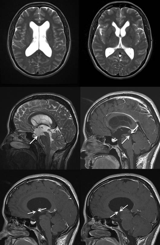
The 43-year-old female patient had complained of progressively worsening headache attacks over several weeks. The MRimaging shows an obstructive hydrocephalus which is caused by a non-enhancing tumor in the area of the aqueduct. The lateral ventricles and the third ventricle are considerably expanded. In the sagittal views one can see the bulging of the floor of the third ventricle of the brain into the prepontine cistern and the compression of the pituitary stalk as an indication of the pressure gradient between the third ventricle and the cisterns. The very narrow foramina of Monro are particularly noticeable. A right precoronal burr hole was chosen as the access point.
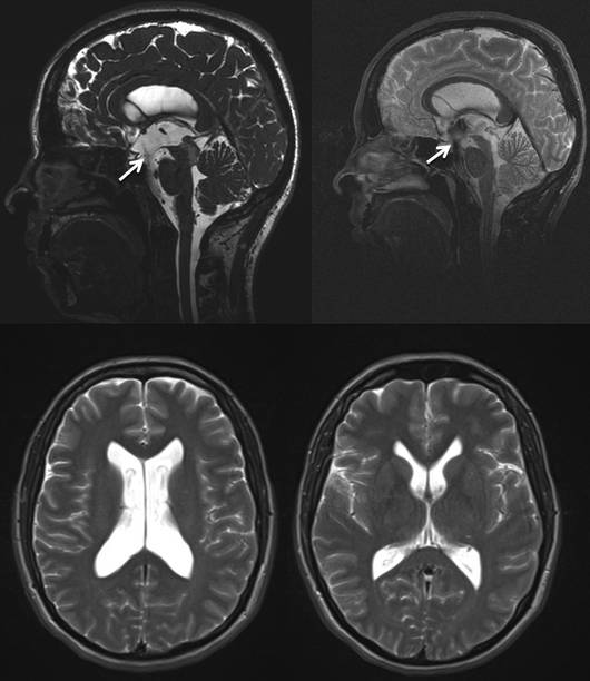
The headache symptoms were alleviated immediately after the operation. The neurological findings are normal. The postoperative MR images show a patent ventriculostomy with strong flow void phenomenon between the prepontine cistern and the third ventricle. The ventricle size has decreased considerably.