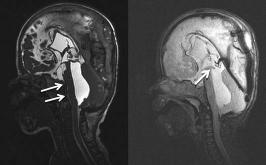
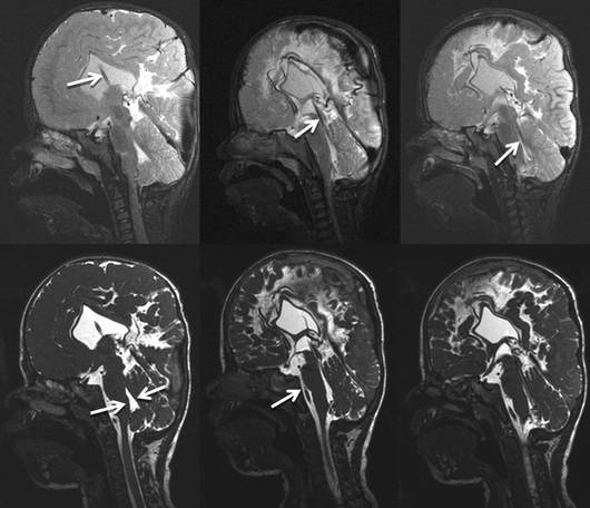
A one-year-old boy who had been born prematurely presented with a posthemorrhagic hydrocephalus. At the age of three months a ventriculoperitoneal shunt was placed. A progressively isolated fourth ventricle then proceeded to develop. The sagittal MR imagesshow a clearly enlarged fourth ventricle with compression of the brain stem against the clivus. The flow-sensitive T2 weighted images show no flow in the aqueductal area. Clinical examination of the boy confirmed psychomotor retardation with considerable coordination disorders.
The postoperative MR images show the correct position of the stent, which now connects the fourth ventricle with the third ventricle and the lateral ventricles. The size of the fourth ventricle is almost normal again. Brain stem compression can no longer be seen. In spite of psychomotor development retardation, the neurological development of the young patient was good following the operation.