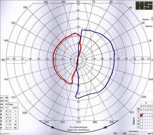
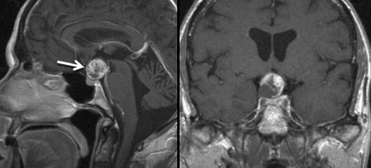
This 57-year-old male presented with severe visual impairment and a narrowed visual field. The ophthalmologic examination recorded visual acuity of 0.1 in the left eye and 0.2 in the right eye as well as severe bitemporal hemianopsia. MR imaging identified the cause of the symptoms as an inhomogeneously enhancing, suprasellar lesion with compression of the optic chiasm and prefixed chiasm. According to MRI criteria, a craniopharyngioma was suspected.
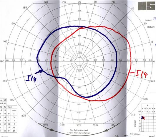
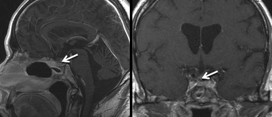
The ophthalmologic deficiencies improved quickly following the operation. Measurement of the visual field 14 days postoperatively already showed a considerable improvement of the outer limits. One year after the operation, the visual field is now only slightly narrowed in the lower quadrant. Visual acuity is 1.0 in the right eye and 0.9 in the left eye. The diabetes insipidus which developed shortly after the operation has also completely disappeared. Due to partial insufficiency of the gonadotropic and thyreotropic axes, the patient has to continue taking testosterone and thyroxine supplements. The adrenocorticotropic axis is intact. MR imaging one year after the operation showed complete removal of the tumor with a preserved pituitary stalk and pituitary gland. Two years after surgery, no recurrence has occurred.