Case description
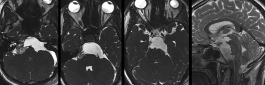
This 32-year-old female presented with headaches and temporary double vision. The MR imaging revealed a largeprepontine epidermoid which has displaced the brainstem dorsally quite considerably.
Pre-operative Images
Video
Result
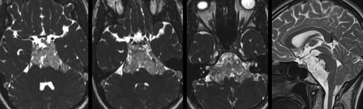
The postoperative MRimages show the extent of the tumor resection. There was only a small residual tumor remaining in the right cerebellopontine angle which was subsequently removed in a second operation via a right retrosigmoid craniotomy. A slight hypesthesia in the left side of the face and a partial abducens palsy which occurred after the second operation, disappeared completely within the first two months after surgery. The patient now displays no neurological deficits and the MR imaging revealed no evidence of a recurrent tumor.
Post-operative Images