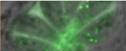

- Visualization and identification of host-pathogen-interactions in the infection models,
Staphylococcus aureus and Toxoplasma gondii
Picture: green fluorescent S. aureus in large vacuoles of a HeLa cell.

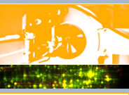
- Subcellular localisation and orientation of proteins by microscopy and high-throughput techniques (topological proteomics).
Picture: Volume reconstruction of an endothelial cell (EaHy926) expressing a yellow-fluorescent plasma membrane marker.
- Data integration and data mining of microarray- and proteomics results via Bioinformatics
Picture: Animation of gene up- and downregulation (red and blue, respectively) on the 14 nuclear chromosomes of the malaria parasite during the intraerythrocytic cycle.
For more information read the open-access article: Scholz M., Fraunholz M.J. (2008) A computational model of gene expression reveals early transcriptional events at the subtelomeric regions of the malaria parasite, Plasmodium falciparum. Genome Biol. 9(5):R88.
- AureusDB: a Staphylococcal genome database (Click on the link to access the database)