Aneurysms are blood-filled bulges of the cerebral arteries. They develop mainly at the bifurcation of blood vessels. They originate in abnormal development (congenital weakness) of the cerebral vascular wall. Under the influence of blood pressure it leads to aneurysmal extension of this weakness of the wall during further life. Approximately 1-2% of the population has such aneurysms. In numerous cases they are clinically completely silent. Due to increasing size they can induce though pressure symptoms in the neighbouring brain parts and brain nerves, and then require treatment. If such an aneurysm breaks under the influence of blood pressure, bleeding in the brain occurs (either as subarachnoid haemorrhage in the subarachnoid cavities or in the cerebral tissue itself, i.e. intracerebral haemorrhage). In about 50% of cases the initial bleeding from the cerebral aneurysm is lethal. Many survivors have severe obstructions. That is why it is important to occlude the aneurysm in order to prevent any further bleeding.
In general the aneurysm manifests itself clinically in one of two ways. In the first it can lead to a rupture of the aneurysm, resulting in a subarachnoid haemorrhage. In the other it can lead to symptomatic malfunctions through the pressure of the ballooning aneurysm on the neighbouring brain structures (e.g. brain nerves). An increasing number of aneurysms are discovered during routine examinations of the brain, e.g. investigation of unspecific headaches or within concussion diagnostics.
The aneurysm diagnostics takes place for subarachnoid haemorrhage by X-ray computed tomography (CT) combined with computed tomography angiography (CTA). Additionally, digital subtraction angiography (DSA) is available. With these examination techniques cerebral vessels are depicted. In the case of asymptomatic aneurysms magnetic resonance imaging (MRI) combined with magnetic resonance angiography (MRA) is applied.
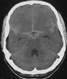
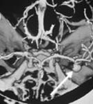
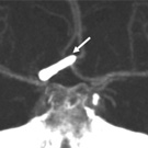
Fig. 1:
A: CT with the image of subarachnoid haemorrhage.
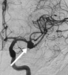
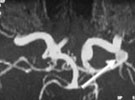
B - D: Image of aneurysm of the left middle cerebral artery made with various techniques. (B: CTA C: DSA D: MRA)
If the aneurysm is an incidental finding, it has to be determined whether treatment is necessary. Whether the aneurysm has to be treated depends first of all on the size of the aneurysm, its location and the age of the patient. An individual consultation is essential and takes place during an interview in our outpatient department. If the presence of an aneurysm manifests through a rupture, i.e. through subarachnoid haemorrhage, there is a life-threatening and immediate need of treatment.
After depicting the aneurysm, therapy takes place aimed at isolating the aneurysm from the blood circulation and thereby preventing (further) rupture (burst). In patients with subarachnoid haemorrhage a surgical intervention is indicated on the day of occurrence of bleeding or on the following day.
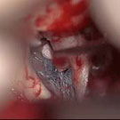
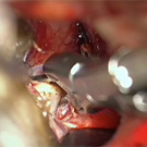
In many aneurysms the therapy of choice is surgical treatment with microsurgical occlusion of the aneurysm neck by attaching a clip (Fig. 1). In some aneurysms the use of an endoscope is very helpful in order to be able to completely view the aneurysm (Fig. 2).
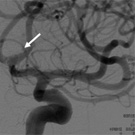
Fig. 2.
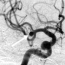
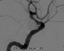
A: DSA with the image of an aneurysm of the middle cerebral artery (arrow).
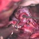
B: The video shows clipping of the aneurysm.
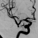
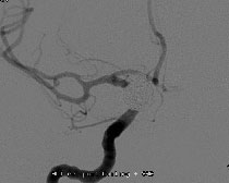
C: The postoperative DSA shows complete occlusion of the aneurysm.
Fig. 3.
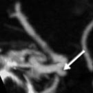
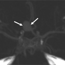
A: CTA with the image of an aneurysm of the right internal carotid artery (arrow).
B: The video shows clipping of the aneurysm and the advantage gained by use of the endoscope in order to see under the artery. Only by using the endoscope can the aneurysm be seen completely.
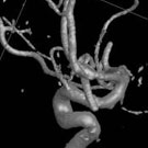
C: Postoperative 3D rotational DSA shows complete occlusion of the aneurysm.
Fig. 4.
A: CTA with the image of an aneurysm of the anterior cerebral artery (anterior communicating artery) (arrows).
B: The video shows clipping of the aneurysm and the use of intraoperative fluorescence angiography that confirms complete occlusion of the aneurysm.
C and D: The postoperative CTA (C) and DSA (D) show complete occlusion of the aneurysm.
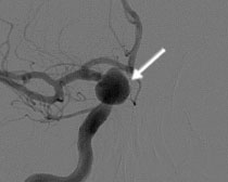
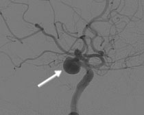
Fig. 5.
A: DSA with the image of a large aneurysm of the right internal carotid artery (arrow).
B: The postoperative DSA shows complete occlusion of the aneurysm by platinum coils (intervention carried out by senior physician Kirsch, Neuroradiology).
|